OCT: a cross-section of the eye
Optical coherence tomography does with light what ultrasound does with sound. In seconds it builds a cross-section of the retina and the optic nerve, in detail finer than a hair's width.
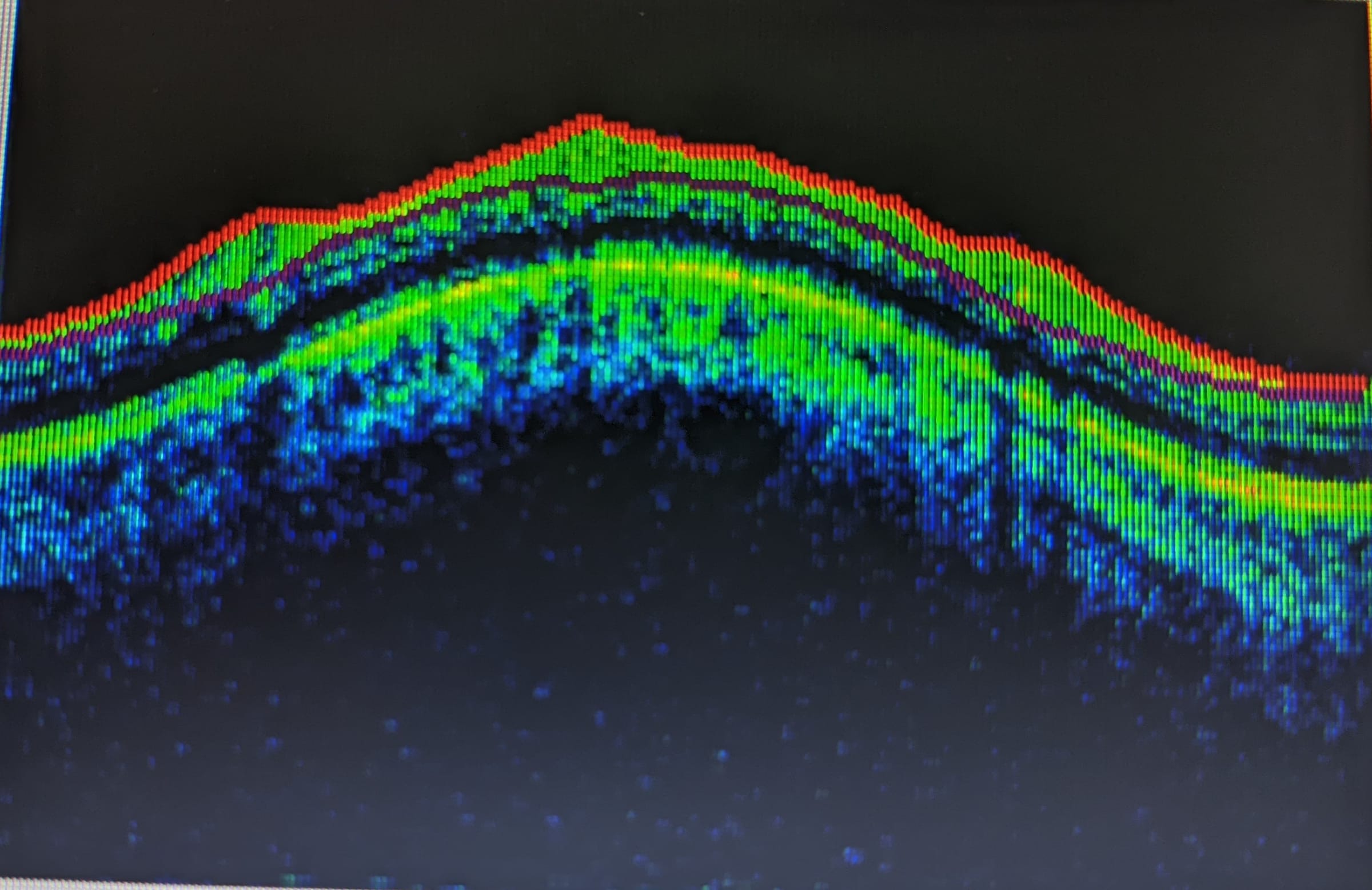
It takes seconds, and nothing touches the eye.
It shows swelling, fluid and thinning before they are felt.
It uses light, so there is no radiation.
How it works
The machine sends a beam of near-infrared light into the eye and measures the light that returns from each layer. Layers that are deeper send their light back a little later, and the machine uses that delay to measure how deep each layer lies. Thousands of these readings, side by side, make the picture, which is why it is called an echo of light.
What it shows
- Swelling and fluid at the macula, in diabetic eye disease and other conditions.
- Holes and membranes at the centre of the retina.
- Thinning of the nerve fibre layer, which glaucoma causes before any sight is lost.
- Whether treatment is working, by comparing scans over time.
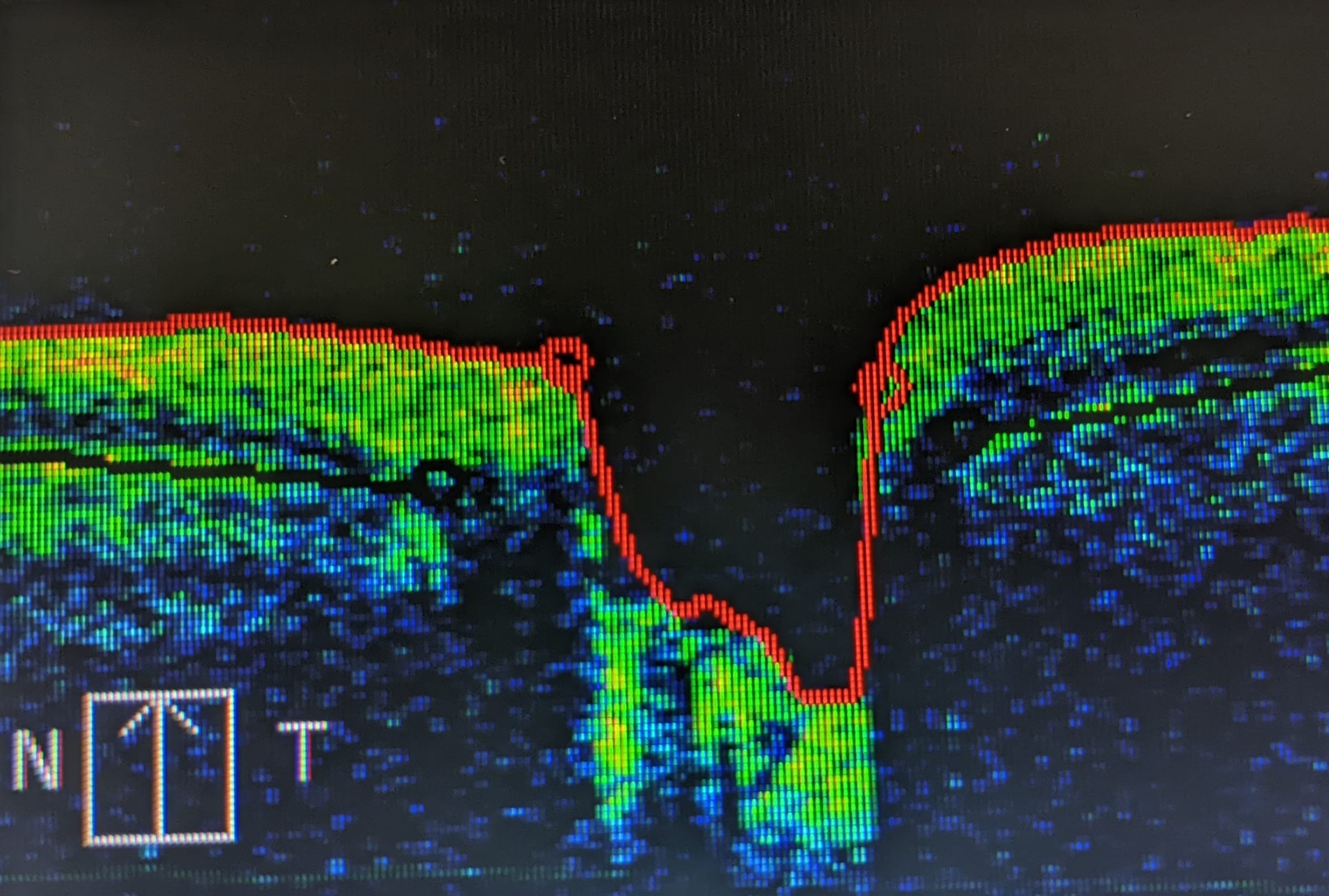
What you do
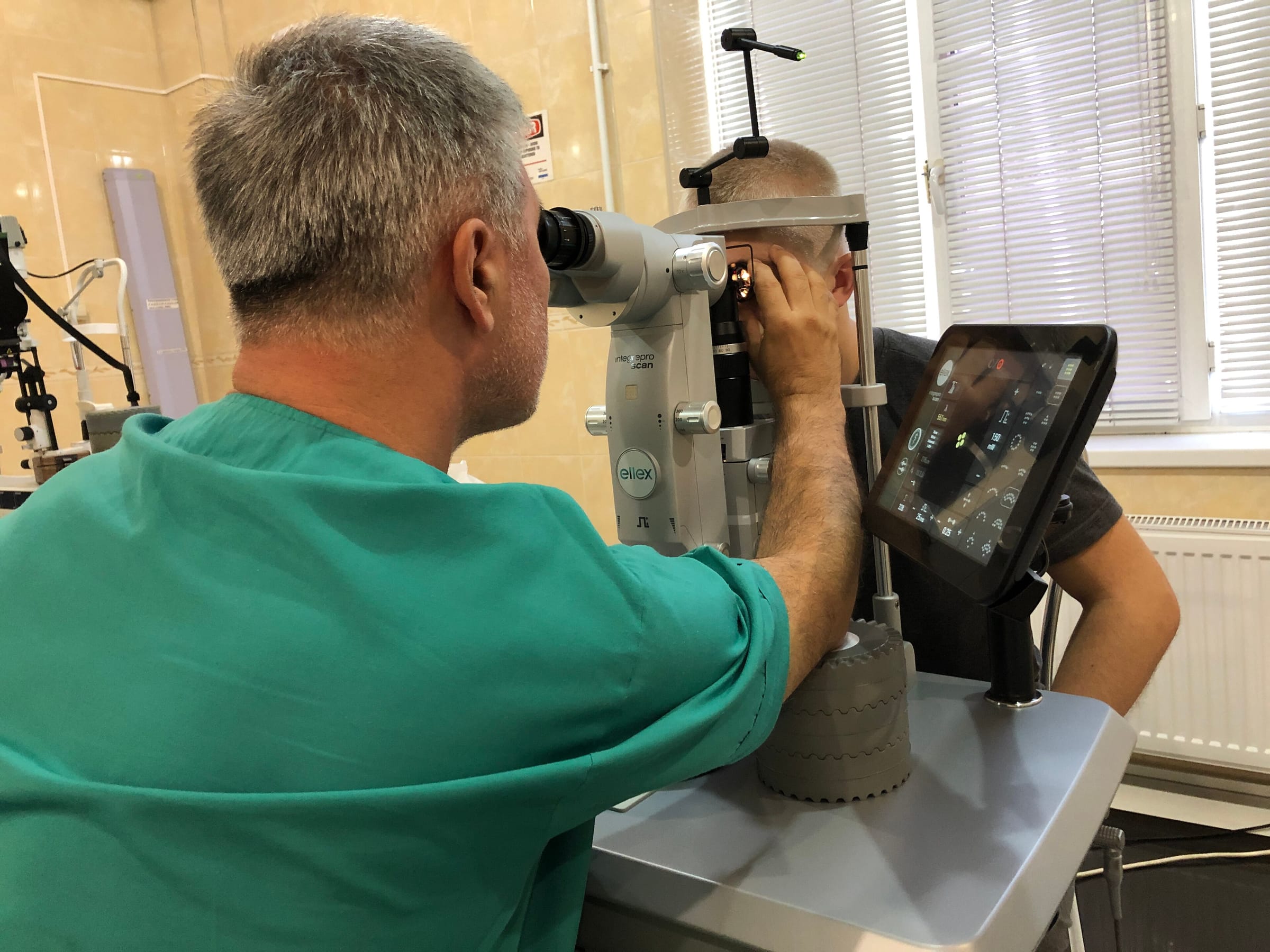
You rest your chin on a support and look at a small light for a few seconds, one eye at a time. Nothing touches your eye. Sometimes drops are used to widen the pupil first.
Keep reading.
Book an eye consultation.
Ask for a time, or leave your number and we will call you.
Or call 1800 210 0108, toll-free.
Quick answers.
Is OCT safe?
Yes. It uses harmless low-power light, with no radiation, and can be repeated as often as needed.
Does it replace the doctor's examination?
No. It adds to it, by showing what cannot be seen from the front.
Pictures not taken by the hospital: Bobjgalindo, CC BY-SA 4.0, Wikimedia Commons.