Corneal mapping
Two tests look closely at the cornea: one maps its shape, the other counts its cells. Surgeons rely on both before they operate.
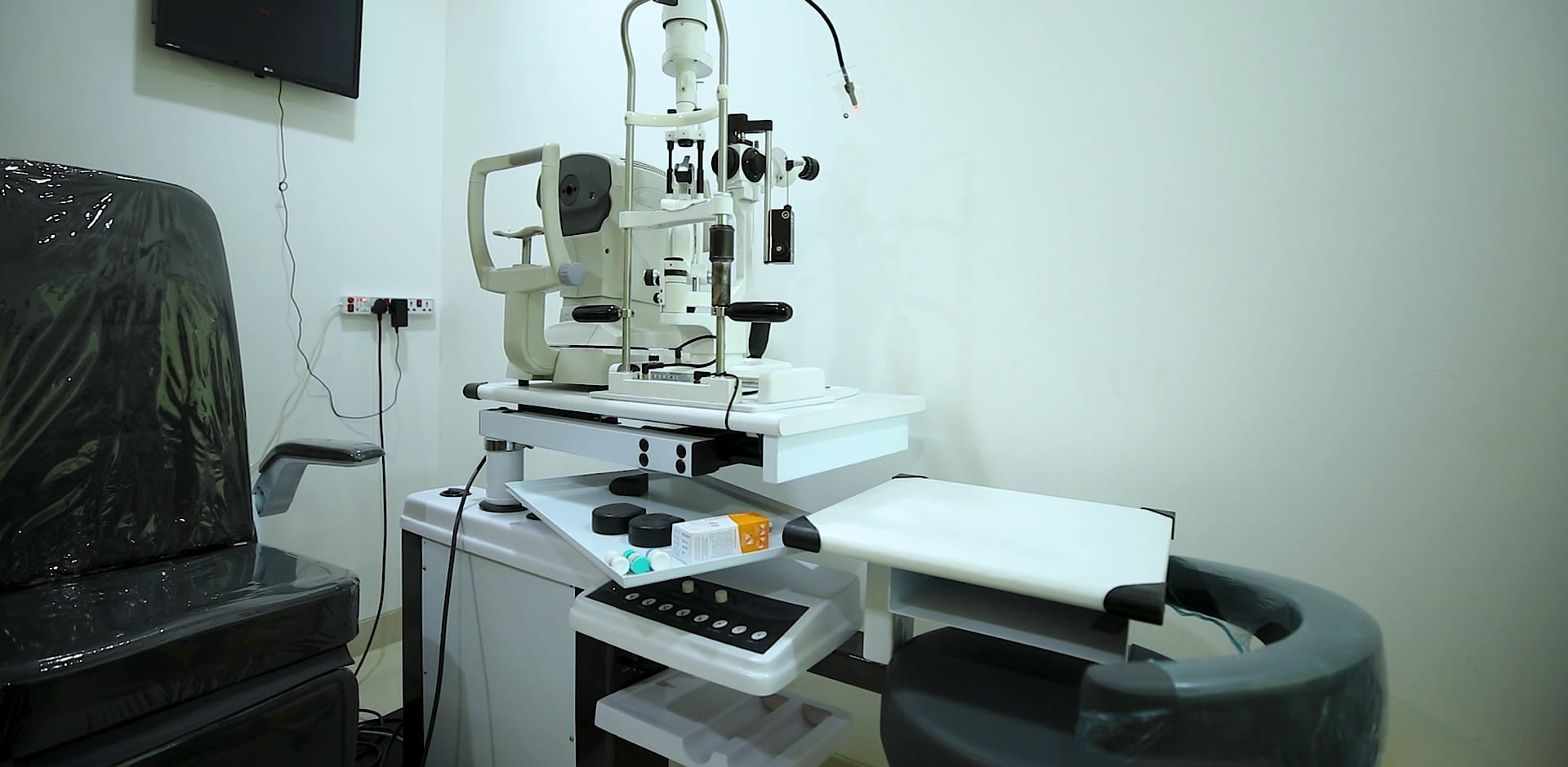
Topography maps the curve of the cornea in thousands of points.
Specular microscopy counts the cells that keep it clear.
Both are done without touching the eye.
Corneal topography
Rings of light are reflected from the front of the eye, and the machine turns their distortion into a colour map of the cornea's curvature. It shows the shape of the eye as no glasses test can.
It finds early keratoconus, measures astigmatism and tells whether a cornea is safe to reshape. It is part of the planning for LASIK, PRK and ICL, and for the power of a toric lens in cataract surgery.
Specular microscopy
The inside of the cornea is lined with a single layer of cells, the endothelium, that pump fluid out and keep the cornea clear. They do not grow back. The specular microscope photographs them without touching the eye, counts them, and shows their size and shape.
A surgeon wants to know this before cataract surgery and before an ICL, and in any eye with a cloudy cornea.
What you do
Rest your chin and look at a ring of light, or a small target, for a moment. There is a bright flash, or a photograph. Painless and contactless.
Keep reading.

Book an eye consultation.
Ask for a time, or leave your number and we will call you.
Or call 1800 210 0108, toll-free.
Quick answers.
Why must I stop wearing contact lenses before the test?
Contact lenses change the shape of the cornea for a while. The map is only true once the cornea has settled.
What if the cell count is low?
Surgery can often still be planned, with more care, or another operation may suit you better. That is the reason for the test.