Digital X-ray
A digital X-ray is the quickest picture of a bone or a chest. It appears on the screen in seconds, at a lower dose than film needed.
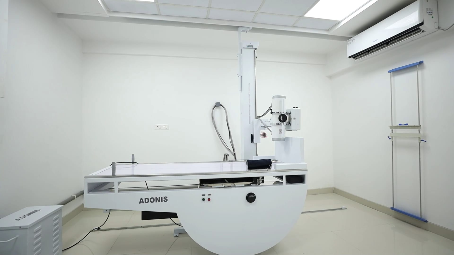
The picture appears in seconds.
A contrast dye shows the gullet, the bowel or the kidneys.
A scanogram measures the whole length of a limb.
How it works
An X-ray tube sends a small beam through the body, and a detector records how much gets through. Bone stops more of the beam than soft tissue, so it shows white. A digital detector, in place of film, gives the picture at once, and it can be adjusted for brightness and contrast.
What it is used for
- A broken bone, or a worn joint.
- An infection in the lung.
- The position of a tube, or of a joint replacement.
- The chest, and the abdomen.
With a dye
Barium
- A swallow, meal or enema outlines the gullet, the stomach or the bowel.
IVP
- The intravenous pyelogram follows a dye through the kidneys and the ureters.
Lower bowel
- Defecography and fistulogram look at the movement and passages of the lower bowel.
Whole limb scanogram
- Records the whole length of a leg or arm on one image.
- Lengths, alignment and deformity can then be measured before a decision on surgery.
Digital X-ray
ADONIS, at Tesla. An ADONIS diagnostic X-ray system, with a digital detector.
- Pictures in seconds.
- Barium studies and IVP.
- Whole limb scanogram.
- A dose lower than film needed.
Keep reading.
Book your scan.
Ask for a time, or leave your number and we will call you.
Or call 1800 210 0108, toll-free.
Quick answers.
Is an X-ray safe?
The dose of a single X-ray is small. It is done when it is needed. Tell us if you might be pregnant.
Do I need to prepare?
For a plain X-ray, no. For a barium study or an IVP, you may be asked to fast or to take a preparation. The desk will tell you when you book.